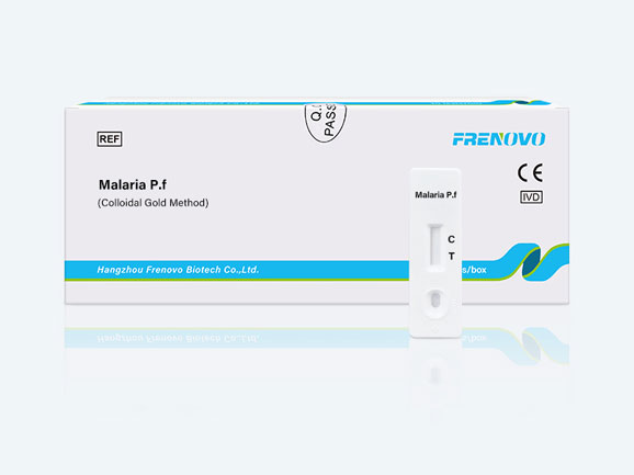

This Malaria Plasmodium falciparum (P.f.) Rapid Test is a qualitative test for the detection of histidine-rich protein 2 antigen (HRP2) of P.f. in human whole blood. This test is for In-Vitro Diagnostic use only.
Malaria is one of the world’s most prevalent parasitic diseases and ranks third in the world among major infectious diseases interms of mortality. The protozoal parasites that cause malaria are from the Plasmodium genus. Four species of Plasmodium protozoa cause malaria: Plasmo-dium falciparum, Plasmodium vivax, Plasmodium malariae and Plasmodium orale. Transmitted principally by the Anopheles mosquito, malaria infections may also occur from contacting infected blood, such as from blood transfusions.
This Plasmodium falciparum (P.f.) malaria test is a rapid, in-vitro immunodiagnostic test for the detection of circulating P.f.antigen in whole blood. The test uses antibodies that are specific for the histidine-rich protein 2 antigen(HRP-2) of P.f.\Whole blood (5 uL) is applied to the sample pad where the red blood cells are lysed with a specially formulated solution. The label pad that is next to the sample pad on the strip is impregnated with blue latex that has an anti- HRP-2 antibody coupled toit. The label pad is also impregnated with purple latex that is coupled to a control antibody. A second anti-HRP-2 antibody is immobilized on the test strip at the test line region. A control material is immobilized on the strip at the control line region.
When a positive sample is applied to the sample pad, P.f. antigen in the sample contacts the latex-labeled antibody and binds to it. A washing reagent is then added to a test vial, and the strip is placed in the vial. As the liquid flows along the length of the strip, any antigen-latex complexes also migrate with the liquid. These complexes are captured by their respective antibodies at the test and control line regions. If a sample contains P.f. antigen, a blue line will form in the test region. If no P.f. antigen is present, a blue line will not form in the test region. A purple control line will always appear in the control region if the test has been properly performed.

